Animal Cell Nucleus
The nucleus is a highly specialized organelle that serves as the information and administrative center of the cell. This organelle has two major functions. It stores the cell's hereditary material, or DNA, and it coordinates the cell's activities, which include intermediary metabolism, growth, protein synthesis, and reproduction (cell division).

Only the cells of advanced organisms, known as eukaryotes, have a nucleus. Generally there is only one nucleus per cell, but there are exceptions such as slime molds and the Siphonales group of algae. Simpler one-celled organisms (prokaryotes), like the bacteria and cyanobacteria, don't have a nucleus. In these organisms, all the cell's information and administrative functions are dispersed throughout the cytoplasm.
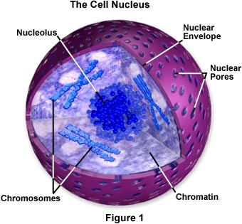
The spherical nucleus occupies about 10 percent of a cell's volume, making it the cell's most prominent feature. Most of the nuclear material consists of chromatin, the unstructured form of the cell's DNA that will organize to form chromosomes during mitosis or cell division. Also inside the nucleus is the nucleolus, an organelle that synthesizes protein-producing macromolecular assemblies called ribosomes.
A double-layered membrane, the nuclear envelope, separates contents of the nucleus from the cellular cytoplasm. The envelope is riddled with holes called nuclear pores that allow specific types and sizes of molecules to pass back and forth between the nucleus and the cytoplasm. It is also attached to a network of tubules, called the endoplasmic reticulum, where protein synthesis occurs. These tubules extend throughout the cell and manufacture the biochemical products that a particular cell type is genetically coded to produce.
Chromatin/Chromosomes - Packed inside the nucleus of every human cell is nearly 6 feet of DNA, which is divided into 46 individual molecules, one for each chromosome and each about 1.5 inches long. Packing all this material into a microscopic cell nucleus is an extraordinary feat of packaging. For DNA to function, it can't be crammed into the nucleus like a ball of string. Instead, it is combined with proteins and organized into a precise, compact structure, a dense string-like fiber called chromatin.
Each DNA strand wraps around groups of small protein molecules called histones, forming a series of bead-like structures, called nucleosomes, connected by the DNA strand. Under the microscope, uncondensed chromatin has a "beads on a string" appearance.
The string of nucleosomes, already compacted by a factor of six, is then coiled into an even denser structure, compacting the DNA by a factor of 40. This compression and structuring of DNA serves several functions. The overall negative charge of the DNA is neutralized by the positive charge of the histone molecules, the DNA takes up much less space, and inactive DNA can be folded into inaccessible locations until it is needed.
There are two types of chromatin. Euchromatin is the genetically active portion and is involved in transcribing RNA to produce proteins used in cell function and growth. Heterochromatin contains inactive DNA and is the portion of chromatin that is most condensed, since it not being used.
Throughout the life of a cell, chromatin fibers take on different forms inside the nucleus. During interphase, when the cell is carrying out its normal functions, the chromatin is dispersed throughout the nucleus in what appears to be a tangle of fibers. This exposes the euchromatin and makes it available for the transcription process.
When the cell enters metaphase and prepares to divide, the chromatin changes dramatically. First, all the chromatin strands make copies of themselves through the process of DNA replication. Then they are compressed to an even greater degree than at interphase, a 10,000-fold compaction, into specialized structures for reproduction, termed chromosomes. As the cell divides to become two cells, the chromosomes separate, giving each cell a complete copy of the genetic information contained in the chromatin.
Nucleolus - The nucleolus is a membrane-less organelle within the nucleus that manufactures ribosomes, the cell's protein-producing structures. Through the microscope, the nucleolus looks like a large dark spot within the nucleus. A nucleus may contain up to four nucleoli, but within each species the number of nucleoli is fixed. After a cell divides, a nucleolus is formed when chromosomes are brought together into nucleolar organizing regions. During cell division, the nucleolus disappears. Some studies suggest that the nucleolus may be involved with cellular aging and, therefore, may affect the aging of an organism.
Nuclear Envelope - The nuclear envelope is a double-layered membrane that encloses the contents of the nucleus during most of the cell's lifecycle. The space between the layers is called the perinuclear space and appears to connect with the rough endoplasmic reticulum. The envelope is perforated with tiny holes called nuclear pores. These pores regulate the passage of molecules between the nucleus and cytoplasm, permitting some to pass through the membrane, but not others. The inner surface has a protein lining called the nuclear lamina, which binds to chromatin and other nuclear components. During mitosis, or cell division, the nuclear envelope disintegrates, but reforms as the two cells complete their formation and the chromatin begins to unravel and disperse.
Nuclear Pores - The nuclear envelope is perforated with holes called nuclear pores. These pores regulate the passage of molecules between the nucleus and cytoplasm, permitting some to pass through the membrane, but not others. Building blocks for building DNA and RNA are allowed into the nucleus as well as molecules that provide the energy for constructing genetic material.
The pores are fully permeable to small molecules up to the size of the smallest proteins, but form a barrier keeping most large molecules out of the nucleus. Some larger proteins, such as histones, are given admittance into the nucleus. Each pore is surrounded by an elaborate protein structure called the nuclear pore complex, which probably selects large molecules for entrance into the nucleus.
