
Java and Flash Tutorial Basics
Interactive Java and Flash tutorials have been developed to help students explore complex concepts in all phases of optical microscopy, the physics of light and color, photomicrography, and digital imaging technology. The tutorials are embedded within web pages that contain accompanying discussions about the subject phenomenon and instructions for use and control of the applets.
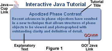
Links to the tutorials appear serially in feature articles, review papers, and general discussions about various topics in microscopy. A typical embedded interactive Java tutorial linking graphic is illustrated in Figure 1. Direct links to index pages for both Java and Flash tutorials appear throughout the microscopy primer.
Linking graphics for interactive tutorials are color-coded to differentiate between those containing Java applets and those featuring Flash-coded programs. The color code for Java applet linking graphics is a light blue-gray background hue (Figure 1) with darker blue text. A banner appearing across the top of the graphic announces the type (Java or Flash) of interactive tutorial and title bar within the graphic indicates the topic of the tutorial (tutorial title). In the central portion of the graphic is explanatory text that describes the applet topic and actions. To launch the page containing a tutorial, use the mouse cursor to click on the upper left or lower right-hand graphic elements in the linking graphic. For Java tutorials, the upper left-hand graphic is an animated coffee cup "button" and the lower right-hand graphic is a Go! Java button (Figure 1). Clicking on either button will launch the applet.
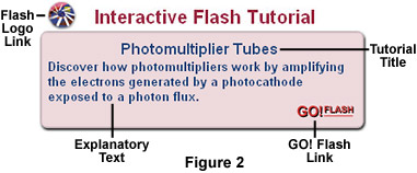
In contrast, linking graphics for interactive Flash tutorials have a separate color code to allow quick differentiation from Java applets (Figure 2). The linking graphic background hue for Flash tutorials is a mauve (pink) hue highlighted with blue text. Other features, such as explanatory text and the tutorial title, are arranged in a manner similar to those for Java tutorials. Launching buttons for the Flash tutorials are a miniature Flash logo (Figure 2; upper left corner) and a Go! Flash button (Figure 2; lower right corner). Like the buttons for Java tutorials, using the mouse cursor to click on either button will launched the web page containing a Flash program.
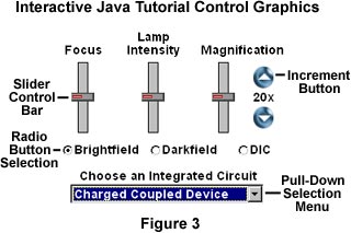
As mentioned above, control of Java and Flash tutorials is accomplished through the utilization of slider bars, radio buttons, and pull-down selection menus. A typical Java control panel is illustrated in Figure 3 for a virtual microscope applet that simulates operation of a microscope. This particular applet has three slider control bars, which are used to adjust specimen focus, illumination intensity, and magnification. To activate a slider bar, place the mouse cursor over the control icon, click the left mouse button, and drag the slider bar either up and down or left and right, depending upon the graphic orientation. As the slider bar is translated, the target algorithm will apply calculations to the image or animation in the applet window. Flash tutorial control panels vary slightly in their look and feel, but operate in a similar manner.
Radio buttons are used to select a new set of conditions for the applet. In the control panel illustrated in Figure 3, the radio buttons are used to toggle between one of three illumination modes for the virtual microscope. When the far-left radio button is selected by clicking with the mouse cursor, Brightfield illumination mode for the virtual microscope is activated and a new palette of images appears in the microscope viewport window (not shown). Likewise, selection of the Darkfield and DIC radio buttons will convert the applet to darkfield and differential interference contrast (DIC) illumination modes, respectively. Radio buttons are used throughout our tutorials in the manner just described to select a particular set of conditions for the Java applet or Flash program.
Many of the tutorials contain a variety of specimens designed to provide comprehensive examples of the phenomenon under study. For instance, the virtual microscope applet discussed above contains a palette of reflected light photomicrographs depicting the surfaces of several integrated circuits (chips). Clicking the mouse cursor on the pull-down menu window will explode a selection palette from which to choose a new image or set of images. When the mouse is moved away from the menu window, the applet starts to download the new images and a message banner appears to warn the user that a short wait will be encountered while the new files are being downloaded.
Use the links provided throughout the microscopy primer to go directly to our specific index pages for interactive Java and Flash tutorials. We hope you enjoy these tutorials and find them useful for exploring new concepts in optical microscopy, photomicrography, and the physics of light and color.
BACK TO MICROSCOPY PRIMER HOME