
Polarized Light Microscopy
Oolite Thin Section in Polarized Light
Oolite, a light gray rock composed of siliceous oolites cemented in compact silica, is formed in the sea. The mineral's name is derived from its structural similarity to fish roe - caviar!
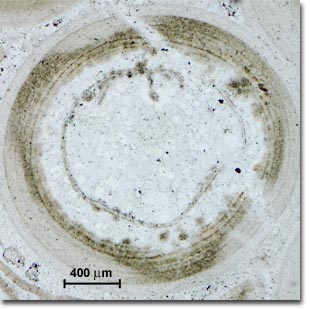
Oolite (above) illuminated with plane-polarized light.
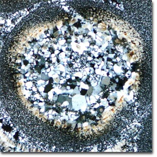
Oolite (above) as imaged through crossed
polarized illumination.
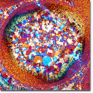
Oolite (above) imaged with crossed polarizers and a
full-wave (first order) retardation plate inserted
between the specimen and analyzer.
Oolite forms in the sea when sand grains are rolled by gentle currents over beds of calcium carbonate or other minerals. These minerals build up around the sand grains and subsequent cementation transforms the grains into coherent rock. The thin sections show the original quartz nuclei on which the build up of carbonate mineral occurred.
In plane-polarized light, the quartz is virtually invisible having the same refractive index as the cement, while the carbonate mineral with a different refractive index shows high contrast. The crossed polarizer image shows quartz grains in greys and whites and the calcium carbonate in the characteristic biscuit colored, high order whites. The groups of quartz grains in some of the cores reveal that these are polycrystalline and are metamorphic quartzite particles. When a full-wave retardation plate is inserted into the optical path, optical path differences become apparent in the specimen, and contrast is enhanced.
Thin sections of the mineral were polished and sealed with a cover glass, then observed and photographed using a Nikon Eclipse E600 microscope equipped with crossed polarizers and an intermediate tube containing an insertable retardation plate. The scale bar represents 400 microns.
Contributing Authors
Phillip C. Robinson - Department of Ceramic Technology, Staffordshire Polytechnic, College Road, Stoke-on-Trent, ST4 2DE United Kingdom.
Michael W. Davidson - National High Magnetic Field Laboratory, 1800 East Paul Dirac Dr., The Florida State University, Tallahassee, Florida, 32310.
BACK TO POLARIZED LIGHT INTRODUCTION