
Polarized Light Microscopy Digital Image Gallery
Trachyte Porphyry
An igneous rock, trachyte primarily consists of alkali feldspars, but also contains plagioclase as well as minor amounts of other minerals, such as olivine, biotite, and pyroxene. It is commonly considered to form via the crystallization and extraction of magnesium, iron, and calcium from a basaltic magma.
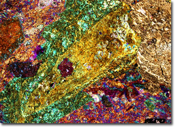
First discovered in ancient Thrace, the name for trachyte is generally believed to have been derived from a Greek word meaning “rough” or “coarse.” Such etymology is quite fitting since trachyte typically occurs as porphyry, a rock in which large, conspicuous crystals are set in a fine-grained matrix. Trachyte also often features a banded or striated appearance due to the flow of the lava from which it formed. The color of the rock varies, but the hues it exhibits are usually light and muted.
Trachyte was a very popular building material in ancient Rome, where it was also utilized for ornamental purposes and to pave roads. The large deposits that the Romans exploited for the material were located on the island of Sardinia. The prevalent use of the rock in the area has resulted in several Sardinian “trachyte towns” where the baths, churches, bridges, arches, tombs, and other notable structures are almost solely comprised of trachyte. Many of these beautiful historical structures can still be seen today.