
Polarized Light Microscopy Digital Image Gallery
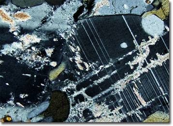
Hornblende Schist
Many significant deposits of valuable gemstones have been found in various schists. Among the most famous are the emerald-rich talc and mica schists that served as “Cleopatra’s Mines” in Upper Egypt near the Red Sea coast, which provided many beautiful pieces of jewelry and ornamental items for the famous queen.
View a second image of Hornblende Schist
Schists are a group of metamorphic rocks that have a visibly foliated crystalline structure. Due to this structure and the platy minerals the rock contains, schists tend to split easily into perfectly thin, flexible sheets along their layers. The minerals that are found in schists vary significantly, but talc, chlorite, muscovite, biotite, and graphite are some of the more common constituents. In order to more explicitly identify samples of the rocks, schists are typically classified and described based upon their mineralogical composition. Hornblende schist, for example, is a variety of schist rich in the amphibole mineral hornblende, though the rock may also contain an abundance of plagioclase feldspar and other substances as well.
Hornblende, like other members of the amphibole group, is a common rock-forming mineral. In fact, hornblende is the most prevalent of all the amphiboles. It is widely distributed in igneous and metamorphic rocks, such as schists. Calcium-rich and monoclinic, hornblende displays the characteristic amphibole structure, which involves four metal sites located between double tetrahedral chains.