Polarized Light Microscopy Digital Image Gallery
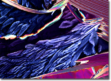
Safranin O
The invention of the compound microscope in the late sixteenth century helped change the way the world is viewed and understood. Though the earliest models were not particularly powerful, they paved the way for improved, higher resolution instruments that were able to reveal tiny details that could hardly have even been imagined before.

View a second image of Safranin O
Not only have improvements to the microscope facilitated numerous discoveries, but the development of a wide array of sample handling and staining techniques has also played an important role in furthering the aims of science. Microscopy stains are often utilized with a wide variety of specimens, especially those that do not readily absorb light. This is because stains render such samples, which could not otherwise be seen, visible to the eye. Stains are also often used in combination with one another in order produce easily distinguishable contrasting colors. For instance, a blue hematoxylin stain might be applied to cell nuclei in order to make the important structures highly discernable from surrounding cytoplasm stained pink with eosin.
Safranin O is a stain frequently utilized in microscopy that appears as dark red to brown crystals at room temperature. According to most sources, safranin O is a mixture of the compounds dimethyl safranin and trimethyl safranin, though some only list the dimethyl compound as a constituent of the stain. Safranin O is most commonly employed for counterstaining nuclei red, but may also be used to stain chromosomes and cell walls. Similar to most substances found in the laboratory, safranin O must be handled with care. The stain can cause both skin and eye irritation, and may be harmful if swallowed, absorbed through the skin, or inhaled. Safranin O is also considered an irritant of mucous membranes and the upper respiratory tract, but is not flammable or explosive under typical laboratory conditions.
