Microscopy Primer
Light and Color
Microscope Basics
Special Techniques
Digital Imaging
Confocal Microscopy
Live-Cell Imaging
Photomicrography
Microscopy Museum
Virtual Microscopy
Fluorescence
Web Resources
License Info
Image Use
Custom Photos
Partners
Site Info
Contact Us
Publications
Home

The Galleries:

Photo Gallery
Silicon Zoo
Pharmaceuticals
Chip Shots
Phytochemicals
DNA Gallery
Microscapes
Vitamins
Amino Acids
Birthstones
Religion Collection
Pesticides
BeerShots
Cocktail Collection
Screen Savers
Win Wallpaper
Mac Wallpaper
Movie Gallery

Polarized Light Microscopy Digital Image Gallery

Margarite

Phyllosilicates with a monoclinic crystal system, a single sheet of mica is comprised of two layers of silicon, each of which exhibits a relative negative charge. These layers are cross-linked together by one of several cations, such as potassium or sodium.

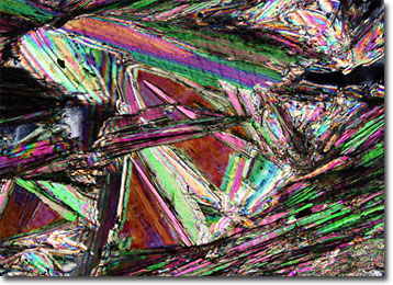
Margarite is a mica that features calcium as the interlayer of cations that holds each sheet of the mineral together. The substitution of this element for those more commonly included in micas results in a less flexible mineral. Thus, although margarite still exhibits the perfect cleavage characteristic to most micas, the thin sheets it produces when cleaved are not as bendable as those of many other varieties. Margarite is, therefore, classified as one of the brittle micas. White, gray, yellowish, or pink in color, margarite typically displays a pearly luster, its name stemming from the Greek word for “pearl.” The mineral generally occurs in low and medium-grade metamorphic rocks, such as mica schists, and often in association the aluminum oxide mineral corundum, which readily weathers to margarite and other aluminous minerals.


BACK TO THE ROCKS AND MINERALS GALLERY

BACK TO THE POLARIZED LIGHT GALLERY

Questions or comments? Send us an email.
© 1998-2022 by Michael W. Davidson and The Florida State University. All Rights Reserved. No images, graphics, scripts, or applets may be reproduced or used in any manner without permission from the copyright holders. Use of this website means you agree to all of the Legal Terms and Conditions set forth by the owners.
This website is maintained by our
Graphics & Web Programming Team
in collaboration with Optical Microscopy at the
National High Magnetic Field Laboratory.
Last modification: Friday, Nov 13, 2015 at 02:19 PM
Access Count Since November 20, 2003: 12220
For more information on microscope manufacturers,
use the buttons below to navigate to their websites: