Polarized Light Microscopy Digital Image Gallery
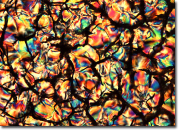
Liquid Crystalline DNA
Deoxyribonucleic acid, better known as DNA, was first discovered in 1869, but its genetic function was not realized until almost 75 year later. About a decade after the comprehension of its true importance, James D. Watson and Francis Crick ascertained the structure of the unusual molecule, which is commonly referred to as a double helix.

View a second, third, fourth, fifth, sixth, and seventh image of Liquid Crystalline DNA
A single molecule of DNA consists of a long chain composed of two strands. Each strand is composed of smaller chemical units called nucleotides, which each include a sugar molecule (deoxyribose), a phosphate group, and one of four different nitrogen-containing compounds called bases. The bases are comprised of adenine (A), cytosine (C), guanine (G), and thymine (T). Each base is capable of forming hydrogen bonds with only one other base; adenine bonds only with thymine and cytosine will only bond with guanine. The nucleotides are held together by covalent bonds between the sugar of one nucleotide and the phosphate of another, while the hydrogen bonds formed between complementary bases are what hold the two strands of a DNA molecule together. This complementarity is also what enables the replication of DNA, ensuring that identical copies of the molecule can be readily reproduced.
DNA is found in all prokaryotic and eukaryotic cells, as well as in a number of viruses. The molecule carries the chemical information that is essential for the direction of protein synthesis and cell replication. In human cells, DNA is wrapped around a complex of histone proteins, forming what is known as chromatin. Chromatin undergoes a process of condensation for cell division or the production of gametes, developing into the higher-order structures called chromosomes. Only one human DNA molecule is present per chromosome. However, many lower types of organisms, such as viruses and bacteria, do not form organized chromosomes at all, though they may still contain DNA.
