
Polarized Light Microscopy Digital Image Gallery
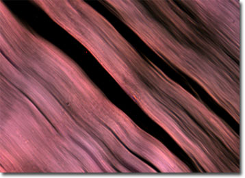
Human Muscle
Muscles are tissues composed of bundles of fibers of varying sizes that can shorten, thicken, or lengthen depending on their location, as well as the message sent to them by controlling neurons. The changes undergone by muscle fibers results in motion, which may be voluntary or involuntary based upon the muscle type.
There are more than 600 muscles in the human body, which collectively comprise about 40 percent of one’s bodyweight. The tissues that constitute these muscles are typically classified, however, as one of three basic types: striated, smooth, or cardiac. Named for its banded appearance under the microscope, striated muscle comprises the bulk of muscle tissue in the human body. Sometimes alternatively known as skeletal muscle, this tissue type is usually attached to the body’s skeleton via fibrous tendons and is under voluntary control. Contrariwise, smooth muscle, which consists of spindle-shaped fibers arranged in sheets along the walls of most hollow organs, and cardiac muscle, which is striated and found only in the heart, are controlled involuntarily by the autonomic nervous system.
Though the movement of striated muscles is voluntary, their muscle tone is involuntarily controlled. The tone of muscles, which are in a constant state of partial contraction, refers to their latent state. Muscles with good tone are firm even when they are relaxed, indicating that they are ready for fast action. Muscles with poor tone, however, are flaccid and take a longer time to react to stimuli because they begin their action from a lesser state of contraction. Pronounced cases of low muscle tone are referred to as hypotonia and in young children may be an indication of hypothyroidism, Down syndrome, or some other condition. Typically, however, those with mild hypotonia can show significant improvement with increased levels of activity.