Polarized Light Microscopy Digital Image Gallery
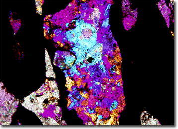
Hematite
The mineral known as hematite derives its name from the Greek word for “blood,” an allusion to its typical red coloring. Relatively hard and heavy, the material is generally composed of approximately 70 percent iron.

Due to its profuseness and high iron content, hematite is considered one of the most important sources of iron ore. The mineral occurs, however, in a variety of forms, some of which are more commercially valuable than others. The type of hematite that occurs in large sedimentary deposits, such as the ones found near Lake Superior and Quebec in North America, is the most notable for its metal ore. Interestingly, according to ancient folklore, such sizable deposits of hematite were formed from the bloodshed of fierce battles seeping into the ground.
In addition to its utilization as a source of iron, hematite is also highly useful to humans as a pigment. Indeed, a form of hematite usually referred to as red ochre occurs in a fine-grained powder, which is often used to impart a reddish hue to paints. All hematite, however, is not red. Some varieties appear a steel gray color and exhibit a beautiful metallic luster. These types of the mineral, which are often called specular iron, specular hematite, or specularite, are sometimes exploited as ornamental stones.
