Microscopy Primer
Light and Color
Microscope Basics
Special Techniques
Digital Imaging
Confocal Microscopy
Live-Cell Imaging
Photomicrography
Microscopy Museum
Virtual Microscopy
Fluorescence
Web Resources
License Info
Image Use
Custom Photos
Partners
Site Info
Contact Us
Publications
Home

The Galleries:

Photo Gallery
Silicon Zoo
Pharmaceuticals
Chip Shots
Phytochemicals
DNA Gallery
Microscapes
Vitamins
Amino Acids
Birthstones
Religion Collection
Pesticides
BeerShots
Cocktail Collection
Screen Savers
Win Wallpaper
Mac Wallpaper
Movie Gallery

Polarized Light Microscopy Digital Image Gallery

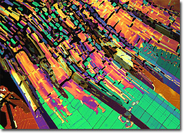
Dicloran

Dicloran is a nitroaniline compound widely utilized as a fungicide. The yellow, crystalline powder is approved for general use in the United States and is sold under a variety of trade names, including Botran, DCNA, Ditranil, and Allisan.
View a second image of Dicloran

A member of the substituted aromatic class of pesticides derived from the parent compound hexachlorobenzene, dicloran is an effective protectant of seeds and ornamental plants. The fungicide may also be utilized on fruit and vegetable crops either before or after harvest to control diseases and may be applied via a variety of methods, including aerial sprays and dusts. Some of the most common items that dicloran is currently utilized to treat are grapes, lettuce, celery, tomatoes, and peaches. The fungicide is sometimes found in formulations with other pesticides, but is incompatible with certain oil-based chemical compounds.

Dicloran is most active against Rhizopus, Botrytis, Monilinia, Sclerotium, and Sclerotinia fungal species. The pesticide’s mechanism of action is believed to be non-specific inhibition of cell division, which facilitates the disruption of nuclear stability. Classified as a class III toxin by the Environmental Protection Agency (EPA), dicloran poses a moderate hazard to fish, birds, and beneficial insects. Studies have also demonstrated that the substance may act as a skin sensitizer and may generate phototoxicity.


BACK TO THE CHEMICAL CRYSTALS GALLERY

BACK TO THE POLARIZED LIGHT GALLERY

Questions or comments? Send us an email.
© 1998-2022 by Michael W. Davidson and The Florida State University. All Rights Reserved. No images, graphics, scripts, or applets may be reproduced or used in any manner without permission from the copyright holders. Use of this website means you agree to all of the Legal Terms and Conditions set forth by the owners.
This website is maintained by our
Graphics & Web Programming Team
in collaboration with Optical Microscopy at the
National High Magnetic Field Laboratory.
Last modification: Friday, Nov 13, 2015 at 02:19 PM
Access Count Since January 23, 2004: 13197
All of the images in this gallery were captured with a QImaging Retiga camera system.
For more information on these cameras, use the button below to access
the QImaging website:
Visit the QImaging website.
For more information on microscope manufacturers,
use the buttons below to navigate to their websites: