Polarized Light Microscopy Digital Image Gallery
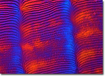
Ctenoid Fish Scale
Scales, which are formed directly in the skin membrane of fish, act as an external form of protection from predators and other dangers in the environment. The number of rows of scales a fish possesses as well as the scale type are characteristics considered in species identification.

Fish scales are typically divided into four main groups, one of which is comprised of ctenoid and cycloid scales. Both of these types of scales are similar in composition, consisting of a rigid surface layer primarily made of crystallized calcium-based salts and a deeper, fibrous layer predominantly composed of collagen. They differ, however, in shape. Cycloid scales feature a smooth posterior edge, but ctenoid scales display ctenii, bony, comb-like structures that decorate the outer margin of the scale. Ctenii may exhibit a wide range of morphologies that vary by species.
Ctenoid scales, which are commonly found in the majority of bony fishes, can sometimes be utilized to estimate the age of the creature to whom they belong. This interesting exploit is possible because when a fish grows, concentric "growth rings" similar to those of trees are formed on its scales. These rings, called circuli, appear closer together when the weather becomes cool and growth slows down, leaving dark bands known as annuli. By counting the annuli, scientists can roughly determine how many winters or cool periods the fish has undergone, resulting in an approximation of its age.
