
Polarized Light Microscopy Digital Image Gallery
Clinochlore
One of the more widely abundant constituents of the chlorite group, clinochlore is a magnesium aluminum silicate hydroxide mineral. Its name was derived from the Greek words for “green” and “inclined,” which are an indication of the mineral’s common color and the pronounced obliqueness of its optical axes.
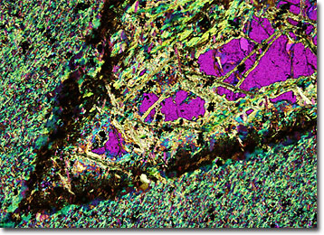
The mineral clinochlore was first identified in 1851 in West Chester, Pennsylvania, but has since been discovered in a number of additional locales in the United States, including New York, Arizona, and New Jersey. Clinochlore can also be found in various other parts of the world, such as Spain, Switzerland, Russia, Turkey, and Italy. Similar to other chlorites, clinochlore exhibits perfect cleavage. The substance is also rather soft and displays a pearly, vitreous luster.
Clinochlore comprises part of a mineral series with chamosite. Though the two are extremely similar, chamosite contains significantly more iron than clinochlore and is more darkly hued, as well as less transparent, than its counterpart. Kaemmererite, on the other hand, is a type of clinochlore that is rich in chromium. This mineral substance is known for the beautiful shades of lavender and crimson that is displays, which stand out vividly against the more common greens of most other chlorites.