
Phase Contrast Image Gallery
Uterine Adenomyosis
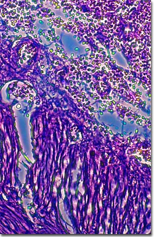
A stained thin section of human uterine tissue exhibiting damage from uterine adenomyosis is illustrated below. As evidenced by the micrograph, combining phase contrast microscopy with classical histological staining techniques in pathological research often yields enhancement of cellular features.
A benign but sometimes painful disease of the uterus, adenomyosis typically affects women between the ages of 40 and 50. Although many cases are asymptomatic, some result in abnormal uterine bleeding and enlargement of the uterus. Tissue that normally lines the interior of the uterus (endometrium) grows into the middle layer of muscles (myometrium), causing blood and debris to collect inside the uterine wall with each progressive menstrual cycle.
The cause of adenomyosis is unknown, but it is believed that the barrier between the endometrium and myometrium, which normally prevents invasion of the endometrial cells into the myometrium, is compromised, allowing the invasion to occur.
The hormone estrogen is probably involved since the symptoms generally disappear with menopause. For most cases, there is little in the way of treatment other than pain management. The only treatment for severe cases is hysterectomy.
Adenomyosis is similar to but not related to endometriosis, in which endometrial cells are found growing outside the uterus altogether.
BACK TO THE PHASE CONTRAST GALLERY