Phase Contrast Image Gallery
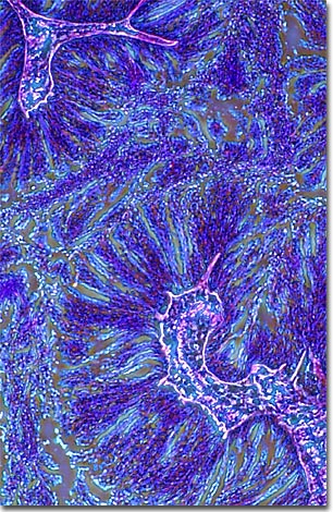
Starfish Testes
A multiply-stained thin section of starfish testes is illustrated below, which was photographed using phase contrast optics. The image was captured using an Olympus IX-70 inverted microscope equipped with a DP-10 digital camera.

Starfish, or sea stars, are perhaps one of the most familiar of marine organisms and are practically a symbol of ocean life. Despite their name, they are echinoderms not fish and breathe through structures on their skin, not through gills.
There are at least 1800 known species of starfish and they occur in all the Earth's oceans (never in freshwater). The greatest variety of species is found in the northern Pacific, from the Puget Sound to the Aleutian Islands. Most starfish have five arms, but a few species have more, as many as fifty arms. These bottom dwellers play a crucial role in the ocean ecosystem, as prey when they are free-floating larvae and as predator when they reach adulthood. Few animals eat adult starfish, which are apparently neither palatable nor nutritious.
Most starfish have two sexes and reproduce by spawning eggs and sperm into the water, but some propagate asexually, at least part of the time. Starfish have the ability to regenerate their arms when lost or injured. Some species take this a step further and clone themselves, separating into pieces, each of which becomes a new animal.
BACK TO THE PHASE CONTRAST GALLERY
