Phase Contrast Image Gallery
Raw Meat
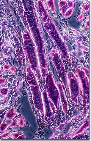
The tissue structure of raw meat is revealed in this phase contrast photomicrograph of a stained thin section of ground hamburger. As evidenced by this micrograph, combining phase contrast microscopy with classical histological staining techniques often yields enhancement of cellular features.

For millions of years, the human diet has included the edible portion of animal tissues, or meat. Meat is an excellent source of protein, providing all nine essential amino acids in addition to vitamins and minerals. There are three types of muscle: smooth, cardiac, and skeletal, but skeletal muscles make up most meats and meat products. Other components include the connective tissue, fat, nerves, and blood vessels that surround and are embedded within the muscles. Lean muscle, regardless of the animal, usually consists of approximately 21 percent protein, 73 percent water, 5 percent fat, and 1 percent ash (the mineral component of muscle).
How animals are handled before, during, and after slaughter affects the structure and biochemistry of the muscles, and therefore, the quality of the meat. After an animal's death, the muscle proteins actin and myosin lose their extendibility and the muscles become stiff, a condition commonly referred to as rigor mortis. Stressing animals just before slaughter and chilling the meat too fast afterwards can result in tough, dry meat. Meat that isn't chilled quickly enough can experience a rapid postmortem pH decline, making it pasty and mushy. Less stressful slaughter techniques and aging the meat in a properly chilled environment produce meats that are more tender and palatable.
VIEW THIS SPECIMEN UNDER FLUORESCENCE ILLUMINATION
BACK TO THE PHASE CONTRAST GALLERY
