Microscopy Primer
Light and Color
Microscope Basics
Special Techniques
Digital Imaging
Confocal Microscopy
Live-Cell Imaging
Photomicrography
Microscopy Museum
Virtual Microscopy
Fluorescence
Web Resources
License Info
Image Use
Custom Photos
Partners
Site Info
Contact Us
Publications
Home

The Galleries:

Photo Gallery
Silicon Zoo
Pharmaceuticals
Chip Shots
Phytochemicals
DNA Gallery
Microscapes
Vitamins
Amino Acids
Birthstones
Religion Collection
Pesticides
BeerShots
Cocktail Collection
Screen Savers
Win Wallpaper
Mac Wallpaper
Movie Gallery

Phase Contrast Image Gallery

Radiolarians

A mixed population of unarranged radiolarian skeletons is illustrated in the photomicrograph below. Medium power objectives with phase contrast optics were used with an Olympus inverted tissue culture microscope to obtain the image.
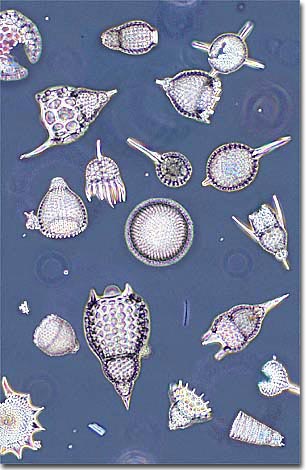
Radiolarians are single-celled protistan marine organisms that distinguish themselves with their unique and intricately detailed glass-like exoskeletons. During their life cycle, radiolarians absorb silica compounds from their aquatic environment and secrete well-defined geometric networks that compose a skeleton commonly known as tests. The radiolarian tests are produced in a wide variety of patterns, but most contain many spines and holes that regulate a network of pseudopods useful in gathering food. Cytoplasm from these microscopic creatures contains numerous vacuoles that fill with air to keep the animals afloat in the upper ocean waters. Radiolarians reproduce asexually, usually by division of the cell (including the exoskeleton), with the remaining daughter cells each regenerating a complete organism. Dead radiolarians accumulate in the ocean floor sediment and are often useful in geological dating experiments.

BACK TO THE PHASE CONTRAST GALLERY

Questions or comments? Send us an email.
© 1998-2022 by Michael W. Davidson and The Florida State University. All Rights Reserved. No images, graphics, scripts, or applets may be reproduced or used in any manner without permission from the copyright holders. Use of this website means you agree to all of the Legal Terms and Conditions set forth by the owners.
This website is maintained by our
Graphics & Web Programming Team
in collaboration with Optical Microscopy at the
National High Magnetic Field Laboratory.
Last modification: Friday, Nov 13, 2015 at 02:19 PM
Access Count Since October 16, 1999: 35191
For more information on microscope manufacturers,
use the buttons below to navigate to their websites: