
Phase Contrast Image Gallery
Obelia Medusa: Second Generation
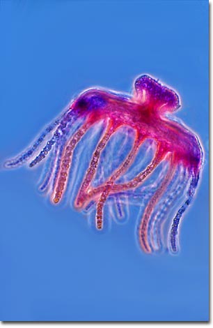
The elegant medusa jellyfish, illustrated in the photomicrograph below, is one of two forms that the hydrozoan Obelia takes during its two-generation life cycle. The free-swimming medusae reproduce sexually, producing eggs and sperm that fertilize to become ciliated, free-swimming larvae (planulae). The planulae remain pelagic for a period of time, eventually attaching to a surface and developing into polyps.
The polyps are stalklike forms that attach to a surface (usually ocean bottom) by means of rootlike filaments. The polyps reproduce asexually, by budding, and create new polyps until it has formed a treelike colony. The colonies are dimorphic, having two types of polyps. Gastrozooids, or hydranths, are the feeding polyps. They have a mouth surrounded by stinging tentacles, giving them a flowerlike appearance, and are responsible for capturing and consuming food. Food is digested in the gastrovascular cavity and provided to the rest of the colony.
Obelia belongs to the phylum Cnidaria, which includes corals, sea anemones, jellyfish, and the freshwater hydra. The many species of this genus are widely distributed throughout all the oceans and are typical of cnidarians, both in their morphology and their life cycle.
BACK TO THE PHASE CONTRAST GALLERY