Phase Contrast Image Gallery
Mammalian Cartilage
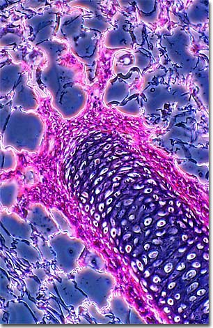
Cartilage is a dense network of collagen fibers, embedded in a firm but plastic-like gelatinous substance, covered by a membrane called the perichondrium. The beautiful image presented below is a phase contrast photomicrograph of a thin section of cartilage that has been stained with eosin and hematoxylin.

The primary function of cartilage in mammals is to form a model for later growth of the bony skeleton, but some parts of the skeleton will retain the cartilage form into adulthood. Unlike bone, cartilage contains no blood vessels. Specialized cells, called chondrocytes, are distributed throughout the collagen gel matrix and obtain nutrition by diffusion through the gel.
Mammals have three types of cartilage. Hyaline cartilage is the type which makes up the embryonic skeleton, most of which will change into bone as the animal matures. In adult humans, it remains unchanged in some parts of the body, including the ends of bones in free-moving joints, at the ends of the ribs, and in the nose, larynx, trachea, and bronchi. Fibrocartilage is a very tough form that is found in the disks of the spinal column. Elastic cartilage makes up the external ear, the auditory tube of the middle ear, and the epiglottis.
BACK TO THE PHASE CONTRAST GALLERY
