
Phase Contrast Image Gallery
Human Scar Tissue
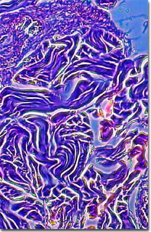
Cut or pierce the skin and the body responds immediately with repair crews of specialized cells for a three-stage rebuilding operation. This results in a scar, or cicatrix in medical terminology. The photomicrograph displayed below is a phase contrast image of a stained thin section of human keloid scar tissue.
First, cells known as phagocytes swarm to the damaged area in an effort to clean up any contaminants and protect the site from infection. New blood vessels begin forming to ensure a blood supply to the new tissue. In the next stage--rebuilding--fibroblasts from the surrounding connective tissue fill the wound with bundles of fibrous connective tissue made up of collagen. During the initial phase, epithelialization, a thin layer of cells grows over the wound, thickening with time. Wound contraction, the next stage, pulls the sides of the wound together. The third and final phase--remodeling--adds more collagen to the scar for added strength, then removes portions of it over time, continually shrinking the scar.
Although the scars are innervated with blood vessels, they lack the oil glands and elastic tissue that normally protect the skin against irritation and can be painful or itchy. When these types of scars cover wide areas of skin, especially from massive wounds or burns, they can make movement difficult as well.
Sometimes abnormally thick scar tissue results from a skin injury and is called hypertrophic scarring. This kind of overscarring can also occur in cases where the healing process doesn't complete the "remodeling" process properly and the wound does not shrink as it should.
Keloids are scars that are excessively thick and fibrous, tumour-like growths that extend beyond the wound's original limits. Keloids seem to have genetic causes and are found primarily in people from African and Asian races.
Although scar reduction and removal is a primary concern in the industrialized world, and one of the primary reasons people see dermatologists, many cultures (including certain subcultures of urban youth) employ deliberate scarring as an ornamentation technique; for aesthetic effect and to indicate status or lineage.
BACK TO THE PHASE CONTRAST GALLERY