
Phase Contrast Image Gallery
House Fly Wings
Insect wings are thought to have evolved from a gill-like thoracic segment present in early insects, which enabled insects to increase the area available for finding food, shelter, and for breeding. The phase contrast photomicrograph below illustrates the veined structure of a typical house fly wing.
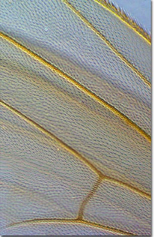
The area between veins consists of a cellular membrane with hair-like projections. Functional wing systems occur only in adult insects with the exception of the Mayfly, so that any insect observed flying can be considered an adult. Many insects are easily identified by their distinctive wing shapes, colors, and patterns.
While at rest, many insects fold their wings, spread them horizontally, or hold them sloped over their backs (house flies), while a few insects wrap their wings around their bodies. Insects have either two or four wings that are used in various ways to coordinate flight, protect the insect, or to attract a mate.
During flight, insects beat their wings rapidly, with house flies having an average repetition of about 200 beats per second. Flies travel at a top speed of four and a half miles per hour, far slower than the dragonfly, which can speed up to 15 miles per hour.
BACK TO THE PHASE CONTRAST GALLERY