Microscopy Primer
Light and Color
Microscope Basics
Special Techniques
Digital Imaging
Confocal Microscopy
Live-Cell Imaging
Photomicrography
Microscopy Museum
Virtual Microscopy
Fluorescence
Web Resources
License Info
Image Use
Custom Photos
Partners
Site Info
Contact Us
Publications
Home

The Galleries:

Photo Gallery
Silicon Zoo
Pharmaceuticals
Chip Shots
Phytochemicals
DNA Gallery
Microscapes
Vitamins
Amino Acids
Birthstones
Religion Collection
Pesticides
BeerShots
Cocktail Collection
Screen Savers
Win Wallpaper
Mac Wallpaper
Movie Gallery

Phase Contrast Image Gallery

Frog Skin

Frogs and the other amphibians, such as toads and salamanders, have unique skin characteristics among vertebrates. A stained thin section of frog skin was photographed using phase contrast optics and is presented below.

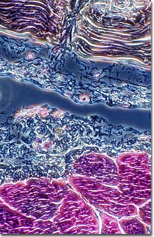
Unlike fish, reptiles, or birds, most amphibians don't have tough, horny scales that function to keep out the elements. Quite the opposite, amphibian skin is delicate and permeable, allowing oxygen and water to pass through its pores. In fact, for many species, the skin is a vital respiratory organ with the underlying dermis richly supplied with blood vessels and lymph spaces.

In order to keep moist, frog skin contains glands that secrete a slimy mucous layer to protect the skin from drying out and help draw in oxygen through the skin. In the water, the mucous secretions help frogs retain a healthy balance of salt and water within their internal tissues. In many species, these glands are modified to produce toxins and other substances that will repel predators. Some frog skin toxins are being researched as potential pain medications.

BACK TO THE PHASE CONTRAST GALLERY

Questions or comments? Send us an email.
© 1998-2022 by Michael W. Davidson and The Florida State University. All Rights Reserved. No images, graphics, scripts, or applets may be reproduced or used in any manner without permission from the copyright holders. Use of this website means you agree to all of the Legal Terms and Conditions set forth by the owners.
This website is maintained by our
Graphics & Web Programming Team
in collaboration with Optical Microscopy at the
National High Magnetic Field Laboratory.
Last modification: Friday, Nov 13, 2015 at 02:19 PM
Access Count Since April 25, 2000: 42198
For more information on microscope manufacturers,
use the buttons below to navigate to their websites: