Microscopy Primer
Light and Color
Microscope Basics
Special Techniques
Digital Imaging
Confocal Microscopy
Live-Cell Imaging
Photomicrography
Microscopy Museum
Virtual Microscopy
Fluorescence
Web Resources
License Info
Image Use
Custom Photos
Partners
Site Info
Contact Us
Publications
Home

The Galleries:

Photo Gallery
Silicon Zoo
Pharmaceuticals
Chip Shots
Phytochemicals
DNA Gallery
Microscapes
Vitamins
Amino Acids
Birthstones
Religion Collection
Pesticides
BeerShots
Cocktail Collection
Screen Savers
Win Wallpaper
Mac Wallpaper
Movie Gallery

Phase Contrast Image Gallery

Fish Skin

A multiply-stained thin section of fish skin is illustrated below, which was photographed using phase contrast optics. The image was captured using an Olympus IX-70 inverted microscope equipped with a DP-10 digital camera.

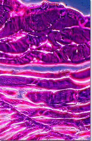
Fish skin or dermis is a key element in fish anatomy and may play a critical role in the swimming dynamics of these aquatic creatures. Composed of collagen fibers arranged in a crossed-helical array, fish skin connects muscle tissue with the bone infrastructure to provide a flexible network that produces an axial undulatory wave motion allowing the fish to propagate through an aqueous environment.

In addition to providing a barrier to the external environment, skin pigment patterns vary widely throughout the fish population, and are effectively utilized to provide camouflage, attract mates, and ward off predators. The outermost layer is composed of hardened, dead keratinized cells composed of scleroprotein with phospholipid side-chains. These thick layers of dead cells probably help to reduce water loss and can add oils, waxes, and lipids to waterproof the skin.

BACK TO THE PHASE CONTRAST GALLERY

Questions or comments? Send us an email.
© 1998-2022 by Michael W. Davidson and The Florida State University. All Rights Reserved. No images, graphics, scripts, or applets may be reproduced or used in any manner without permission from the copyright holders. Use of this website means you agree to all of the Legal Terms and Conditions set forth by the owners.
This website is maintained by our
Graphics & Web Programming Team
in collaboration with Optical Microscopy at the
National High Magnetic Field Laboratory.
Last modification: Friday, Nov 13, 2015 at 02:19 PM
Access Count Since April 23, 2000: 30004
For more information on microscope manufacturers,
use the buttons below to navigate to their websites: