
Phase Contrast Image Gallery
Earthworm Testes and Ovaries
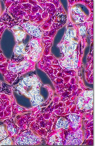
Earthworms, commonly known as night crawlers, are simultaneous hermaphrodites, with each worm having complete male and female reproductive systems that include both testes and ovaries. The photomicrograph below is a phase contrast image of earthworm reproductive tissue stained with a mixture of eosin and hematoxylin.
Although earthworms possess ovaries and testes, they have a protective mechanism against self fertilization and can only function as a single sex at one time. Sexual reproduction occurs when two worms meet and exchange gametes, copulating on damp, wet nights during warm seasons. Fertilized eggs are protected by a cocoon, which is buried on or near the surface of the ground.
Young earthworms are faithful, but miniature, replicas of the parents and become sexually mature in about three months. Earthworms have the ability to regenerate a portion of the body when it is removed or damaged. If the posterior (tail) section of a worm is removed, then either a head or tail is regenerated. However, if a head grows back instead of the original tail, then the double-headed worm will starve. Anterior regeneration always results in a tail section being reproduced. It is interesting to note that portions of several worms can be united to make a single long worm.
BACK TO THE PHASE CONTRAST GALLERY