
Phase Contrast Image Gallery
Cooked Meat
Humans and their ancestors have hunted, killed, and eaten meat for millions of years, but it probably wasn't until the past one million years that meat was cooked before it was consumed. Homo erectus, thought to be the immediate ancestor of Homo sapiens, was the first hominid known to have used fire and presumably became the first to throw their meat "on the barbie."
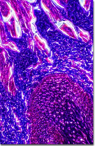
The photomicrograph above illustrates a stained thin section of cooked hamburger meat, one of our favorite staples, using phase contrast optics. Cooking meat makes it safer to eat and causes dramatic changes in its structure and chemistry, many of which make meat easier to chew and more palatable. This is primarily the result of protein denaturation, the physical unfolding of proteins in response to heat. Denaturation causes the change in color that usually occurs when meat is cooked. For example, when it reaches temperatures of 180ş Fahrenheit (82ş Celsius) beef myoglobin, or muscle protein, changes from deep red to a pale gray or brown. The denaturation of the myoglobin proteins make them unable to bind oxygen, which causes the loss of red color. Muscle fibers shorten and toughen as they are heated and eventually their structure breaks down completely. Collagen, or connective tissue protein, also undergoes changes at temperatures above 160ş F (71ş C) that cause the complete denaturation of collagen into a gelatin-like consistency.
BACK TO THE PHASE CONTRAST GALLERY