
Phase Contrast Image Gallery
Cabbage Club Root
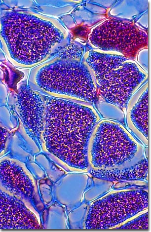
Wilted, yellowing leaves and stunted plants can often signal an infection of club root in cruciferous plants such as cabbage, cauliflower and broccoli, causing significant economic losses to farmers. As evidenced by this micrograph, combining phase contrast microscopy with classical histological staining techniques often yields enhancement of cellular features.
The most distinctive symptom, however, is the abnormal enlargement, or clubbing, of roots where the disease process is taking place. Clubs may cover only sections of roots or they may cover the entire root system of the plant. In some cases, plants may not exhibit any symptoms save for a few small clusters of clubs on the roots.
Club root is caused by the parasitic slime mold, Plasmodiophora brassicae. Slime molds are a primitive group of fungi that are typically found in moist, decaying leaf litter and logs in the forest. Most consume bacteria, yeast, molds, and fungi, but the Plasmodiophorans are obligate parasites and live by consuming the root tissue of plants in the mustard family.
Primary infection occurs when motile spores, known as zoospores, are released into the soil. These zoospores invade plant root hairs and develop into a plasmodium, a structure unique to the slime molds. This spreads through host cells and, in the case of Plasmodiophora, produces the root clubs. The plasmodium is characterized as a slimy, amoeboid-shaped mass of protoplasm with many nuclei, essentially one giant multinucleated cell. As the plasmodium feeds, it enlarges and the nuclei divide about once every 24 hours. The plasmodium may become very large, with millions of nuclei, but when conditions are right, it forms resting spores, which are released into the soil as the host tissue decays. Resting spores of Plasmodiophora can survive in soil indefinitely.
BACK TO THE PHASE CONTRAST GALLERY