
Phase Contrast Image Gallery
Bronchiogenic Carcinoma
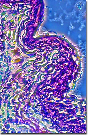
A stained thin section of human lung tissue exhibiting damage from bronchiogenic carcinoma is illustrated below. As evidenced by the micrograph, combining phase contrast microscopy with classical histological staining techniques in pathological research often yields enhancement of cellular features.
Bronchiogenic carcinoma, or lung cancer, is the leading cause of cancer deaths in the United States and most of the developed world. In the 1990s, more women were dying of lung cancer than of breast cancer, which historically had been the major cancer killer in women. Although its cause has been traced to many environmental factors, such as asbestos, benzene, industrial chemicals, and radon exposure, most lung cancers (80-90 percent) are associated with cigarette smoking.
There are two types of lung cancers: non-small cell carcinoma and small cell carcinoma, or oat-cell carcinoma. Of the two, the oat-cell carcinoma is the most aggressive and accounts for about 20-25% of all cases. There are three types of tumors that characterize non-small cell lung cancers: squamous-cell carcinomas, adenocarcinomas, and large-cell carcinomas.
Until the 1900s, bronchiogenic carcinoma, or lung cancer, was a relatively unknown disease. By the 1940s, the incidence of lung cancers and deaths from lung cancer had risen precipitously. At least one physician, Ernst L. Wynder, noticed the association between smoking and lung cancer. In the late 1940s, while still a medical student, he observed an autopsy of a two-pack-a-day smoker and lung cancer patient. Upon interviewing a group of other lung cancer patients, he became suspicious. After his graduation from medical school, he did a study with laboratory mice and became the first to establish a scientific link between cigarette smoking and lung cancer.
BACK TO THE PHASE CONTRAST GALLERY