
Phase Contrast Image Gallery
Black Lung Disease (Anthracosis)
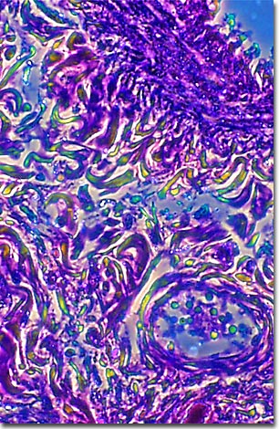
A stained thin section of human lung tissue afflicted with anthracosis, or black lung disease, is illustrated in the photomicrograph presented below. As evidenced by the micrograph, combining phase contrast microscopy with classical histological staining techniques in pathological research often yields enhancement of cellular features.
Black Lung Disease is a form of pneumoconiosis, a condition caused by the habitual inhalation of irritants, coal dust in this case. It is a slow-acting, chronic disease that takes one or two decades to cause symptoms, but its effects can be severely disabling, causing pulmonary emphysema or chronic bronchitis. Cigarette smoking is known to make the symptoms worse.
Anthracosis is the early stage of Black Lung and may be asymptomatic. Early symptoms of the disease include shortness of breath, labored breathing, coughing, and production of phlegm. At this stage, avoiding exposure to coal dust can stop the disease.
Black lung sufferers are at higher risk of developing other potentially fatal diseases such as cancer, tuberculosis, or heart disease. About 4.5 percent of coal miners are affected by black lung. Between 1979 and 1996, 14,156 people died of the disease. Black Lung is probably the most commonly known occupational illness in the United States.
BACK TO THE PHASE CONTRAST GALLERY