Phase Contrast Image Gallery
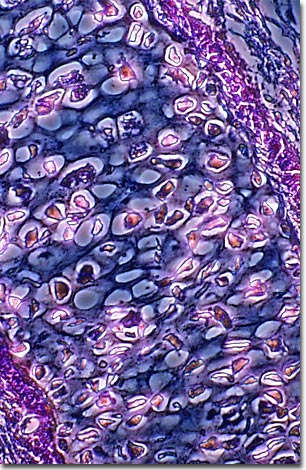
Bird Skin (Epidermis)
A stained thin section of bird skin (epidermis) is illustrated below. As evidenced by this micrograph, combining phase contrast microscopy with classical histological staining techniques often yields enhancement of cellular features.

Birds have a thin and delicate epidermis, or skin, compared to other vertebrates. Their skin produces specialized structures called feathers, which is one of the unique characteristics of birds. Feathers are made up of keratin, a flexible protein that also forms the hair and fingernails of mammals. Thick epidermal scales, like those found in reptiles, usually cover exposed areas of skin, such as the legs and feet.
Birds are found all around the world and in many different habitats, from deserts and savannas to aquatic and marine, from tropical jungles to Antarctic ice fields. Their ability to exist in a wide variety of conditions is due primarily to their highly specialized and protective epidermis and plumage. Over the past 100 million years, feathers have evolved a variety of forms that allow birds to exist in specific environments.
Birds are an ancient lineage dating back to the Cretaceous period, when dinosaurs were at their peak. Scientists are still debating whether birds are dinosaurs that survived the devastation of the asteroid impact 65 million years ago, or if they are simply close relatives of the dinosaurs and arose from a different reptilian ancestor.
VIEW THIS SPECIMEN UNDER FLUORESCENCE ILLUMINATION
BACK TO THE PHASE CONTRAST GALLERY
