Fluorescence Digital Image Gallery
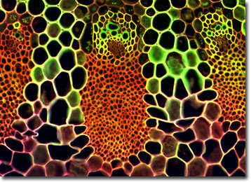
Wheat Tissue Thin Section
Wheat is the common name for any of the cereal grasses belonging to the genus Triticum and is an important food source for people around the world. Evidence shows that wheat grew as a wild grass in the Middle East nearly 10,000 years ago and was in cultivation by 6,000 BC.

The Egyptians were the first to discover how to make yeast-leavened breads, sometime between 3,000 and 2,000 BC. Since wheat is the only grain with enough gluten to make a raised or leavened loaf of bread, it was quickly favored over other grains grown at the time, such as oats, millet, rice, and barley.
The wheat plant is a tall annual and typically grows to a height of four feet. The leaves, similar to those of other grasses, appear early and are followed by slender stalks that produce flowers. Flowers that are fertilized produce seeds called grains or kernels. The wheat kernel is made up of three parts: the endosperm, the bran, and the germ. The tiny germ is the part of the kernel that will sprout and grow into a new wheat plant if the seed is planted.
The most common grain cereal today, world wheat production was more than 590 million metric tons by 1990, an increase of about 30 percent over the average for the period 1979 to 1981. The former Soviet Union was once the world's leading producer, with a near-record 235 million metric tons, but as the central government broke up in 1991, production fell. China was in second place in 1990, the United States in third. Other major wheat producers are India, Canada, France, and Australia. Of the 42 wheat-growing states in the United States, the leading producers are North Dakota, Kansas, Montana, and Oklahoma. In Canada, Saskatchewan, Alberta, and Manitoba are the primary wheat producers.
The specimen presented here was imaged with a Nikon Eclipse E600 microscope operating with fluorite and/or apochromatic objectives and vertical illuminator equipped with a mercury arc lamp. Specimens were illuminated through Nikon dichromatic filter blocks containing interference filters and a dichroic mirror and imaged with standard epi-fluorescence techniques. Specific filters for the wheat tissue stained thin section were a B-2E/C and a Y-2E/C. Photomicrographs were captured with an Optronics MagnaFire digital camera system coupled to the microscope with a lens-free C-mount adapter.
BACK TO THE FLUORESCENCE DIGITAL IMAGE GALLERY
