
Fluorescence Digital Image Gallery
Rhizopus Rot
Rhizopus rot is a soft rot of harvested or over-ripe stone fruits, such as peaches, nectarines, sweet cherries, and plums. Mold species belonging to the genus Rhizopus cause the rot, which initially appears on the fruit as a fuzzy white mass called the mycelium.
The fungus produces enzymes that deteriorate the tissue holding the skin to the flesh of the fruit. Later, it turns dark gray to black as the fungus begins to develop sporangia, the fruiting structures that produce spores. Once released into the air, the spores can infect injured fruit in both orchards and on kitchen countertops. R. stolonifer is one species that commonly affects fruit in household kitchens.
Uninjured fruit are not susceptible to the disease. However, fungal spores can remain dormant until the fruit is harvested. After the fruit ripens and is harvested, the fungus that is already present on the fruit skin is ready to germinate and grow as the fruit ages and the integrity of the skin deteriorates.
Rhizopus is not limited to fruit. It is a highly contagious fungus that can grow even in a refrigerator if food items are not kept covered. Initially the infection begins on one piece of fruit. The fruit quickly becomes covered in sporangia, which eventually rupture, releasing their spores into the air. Any uncovered food in the refrigerator is then vulnerable to contamination by the spores. Green fruit has a better chance of survival since it is inherently more resistant to the disease.
This fungus can also infest many crops and cause root rot when there is a combination of too much soil moisture and damage from insect pests. When the leaves of sugar beets are chewed on by the armyworm (actually the larva of the moth, Pseudaletia unipuncta), the damage creates a point of entry for Rhizopus spores that invade the plant. The fungus grows throughout the plant, but ends up rotting the root.
Not all of the food spoilage caused by this mold is bad. One species, R. oligosporous, has proven economically beneficial to many Asian countries. Added to cooked soybeans, it transforms them into tempeh. The mycelium of the mold grows through the soybean paste holding it together to give tempeh its characteristic chewy texture; fermentation gives it a distinctive flavor. Tempeh is commonly used as a meat substitute and can be deep-fried, sautéed, baked, or steamed.
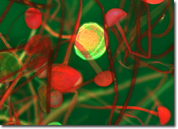
The specimen presented here was imaged with a Nikon Eclipse E600 microscope operating with fluorite and/or apochromatic objectives and vertical illuminator equipped with a mercury arc lamp. Specimens were illuminated through Nikon dichromatic filter blocks containing interference filters and a dichroic mirror and imaged with standard epi-fluorescence techniques. Specific filters for the rhizopus rot stained thin section were a a B-2E/C and a Y-2E/C. Photomicrographs were captured with an Optronics MagnaFire digital camera system coupled to the microscope with a lens-free C-mount adapter.
BACK TO THE FLUORESCENCE DIGITAL IMAGE GALLERY