
Fluorescence Digital Image Gallery
Peach Leaf Curl
Peach leaf curl is a common disease of peach and nectarine trees caused by the fungus Taphrina deformans. Spores of the fungus overwinter on the surface of peach twigs. In spring, the spores multiply during periods of moist weather until the leaf buds swell and open.
Rain is necessary for infection, carrying the spores on a thin film of water into the buds, where leaves are infected. After the deformed and discolored leaves turn brown and fall, they produce powdery gray spores. These are blown by winds to peach twig surfaces and remain there for the winter.
As its name implies, the most common and striking symptom of leaf curl occurs on the foliage. Infected leaves are severely deformed and often display a variety of colors ranging from light green and yellow to shades of red and purple. The fungus causes cells at leaf margins to reproduce quickly and randomly, resulting in leaves that are wrinkled, puckered, and curled. As these infected leaves mature, the fungus produces spores, giving them a dusty appearance, after which the leaves turn brown, shrivel, and drop from the tree. The fruit can also be affected by the fungus, sometimes falling off the tree prematurely, growing in abnormal shapes, or developing wart-like deformities on the surface.
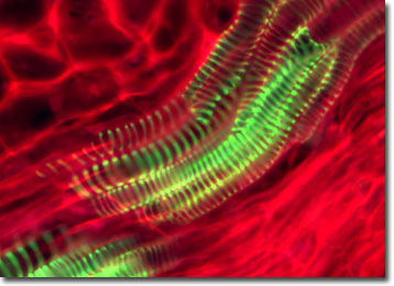
The specimen presented here was imaged with a Nikon Eclipse E600 microscope operating with fluorite and/or apochromatic objectives and vertical illuminator equipped with a mercury arc lamp. Specimens were illuminated through Nikon dichromatic filter blocks containing interference filters and a dichroic mirror and imaged with standard epi-fluorescence techniques. Specific filters for the peach leaf curl stained thin section were a B-2E/C and a Y-2E/C. Photomicrographs were captured with an Optronics MagnaFire digital camera system coupled to the microscope with a lens-free C-mount adapter.
BACK TO THE FLUORESCENCE DIGITAL IMAGE GALLERY