
Fluorescence Digital Image Gallery
Oleander Leaf
Oleander, scientifically described as Nerium oleander, is an ornamental evergreen that belongs to the dogbane family, Apocynaceae. The best-known oleander shrub, called rosebay, is native to the Mediterranean and Middle East regions and is distinguished by dark green leaves that are thick, leathery, and lance-like.
Beginning in spring and continuing through autumn, oleander flowers burst into beautiful rose-like clusters of red, apricot, pink, or white. Common oleander is often found cultivated in greenhouses and is widely grown outdoors in warm regions, where it is often used as a freeway median divider. An adaptable root system allows the plant to root in many different types of terrain including unstable and pebbly ground.
Oleander is beautiful, but extremely deadly. A single leaf can kill an average size adult. In fact, all parts of the plant are highly toxic if ingested. The plant contains a poisonous glycoside, a milky substance that is rich in salicine and other alkaloids. This poisonous sap can paralyze the hearts of humans and animals.
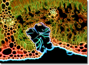
The specimen presented here was imaged with a Nikon Eclipse E600 microscope operating with fluorite and/or apochromatic objectives and vertical illuminator equipped with a mercury arc lamp. Specimens were illuminated through Nikon dichromatic filter blocks containing interference filters and a dichroic mirror and imaged with standard epi-fluorescence techniques. Specific filters for the oleander leaf stained thin section were a UV-2E/C, B-2E/C, and a Y-2E/C. Photomicrographs were captured with an Optronics MagnaFire digital camera system coupled to the microscope with a lens-free C-mount adapter.
BACK TO THE FLUORESCENCE DIGITAL IMAGE GALLERY