Fluorescence Digital Image Gallery
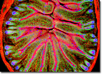
Mouse Intestine Thick Section
Mouse intestines are very much like those of other vertebrate animals. The large intestine is wider and shorter than the small intestine and its primary function is to absorb water and electrolytes from digestive residues and store fecal matter.

The word "mouse" has no meaning in scientific classification, but species of many families of small rat-like rodents are commonly referred to as mice. Most mouse species, such as the common house mouse, belong to the family Muridae. The other major mouse families are Cricetidae (grasshopper mice, harvest mice), Heteromyidae (pocket mice), and Zapodidae (jumping mice and birch mice).
Mice occur in nearly every habitat and on every continent but Antarctica, either naturally or through introduction by humans. Their diet consists of grains, roots, fruits, insects, grass, and "people food". The brownish-gray house mouse originated on the Eurasian continent but gained a worldwide distribution thanks to human dispersal and travel. These mice can multiply quickly, usually breeding every 10 to 17 weeks a year and producing 5 to 10 young per litter.
Laboratory mice are special breeds of house mice and are used in many scientific experiments because of their close mammalian relationship to humans. Compared to larger mammals, mice and other rodents are small, easy to handle, inexpensive to house, and breed quickly. During the late twentieth century (and on into the current century), scientists bred different strains of mice with genetic deficiencies in order to produce models for human diseases.
The specimen presented here was stained with a combination of Alexa Fluor 350 WGA, Alexa Fluor 586 phalloidin, and SYTOX green, then imaged with a Nikon E600 microscope operating with fluorite and/or apochromatic objectives and vertical illuminator equipped with a mercury arc lamp. Specimens were illuminated through Nikon dichromatic filter blocks containing interference filters and a dichroic mirror and imaged with standard epi-fluorescence techniques. The specific filter set for the mouse intestine stained thin section was a DAPI, FITC, Texas Red combination. Photomicrographs were captured with a Optronics MagnaFire digital camera system coupled to the microscope with a lens-free C-mount adapter.
BACK TO THE FLUORESCENCE DIGITAL IMAGE GALLERY
