
Fluorescence Digital Image Gallery
Mite
There are over 20,000 species of mites, tiny arthropod invertebrates belonging to the class Arachnida. Along with the tick, they make up the order Acarina. Mites can be found worldwide in diverse habitats, including brackish water, fresh water, hot springs, soil, on plants and mosses, as well as upon and inside animals. Parasitic forms may live in the nasal passages, lungs, stomach, or even deeper body tissues.
Mites eat plant or animal substances and decaying organisms or parasitize other arthropods, mollusks, and invertebrates. Others can be found on the skin of birds and reptiles, and in the respiratory channels of birds and mammals. Some mites, such as the red spider, harbor diseases. Despite its name, the red spider is a mite, not a spider, which transmits a viral disease that is very damaging to crops. Scab mites, of the family Psoroptidae, can cause skin irritation in humans and fur-bearing animals. Itch mites, members of Sarcoptidae, burrow into the layers of skin of humans, dogs, pigs, goats, and sheep, causing an itchy rash.
Dust mites are tiny arthropods that can be found almost everywhere. They live in house dust and their excrement is the number one indoor allergen. A protein in the fecal products and disintegrating body parts of these miniature mites is the substance that adversely affects those who are sensitive to it. Dust mites are impossible to completely eliminate from living quarters. They have sticky pads on their legs that enable them to burrow into many household items, like furniture, and resist even the most powerful vacuum cleaners.
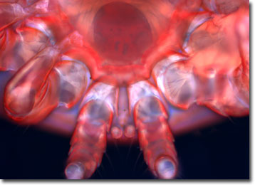
The specimen presented here was imaged with a Nikon Eclipse E600 microscope operating with fluorite and/or apochromatic objectives and vertical illuminator equipped with a mercury arc lamp. Specimens were illuminated through Nikon dichromatic filter blocks containing interference filters and a dichroic mirror and imaged with standard epi-fluorescence techniques. Specific filters for the mite specimen were a UV-2E/C, B-2E/C, and a Y-2E/C. Photomicrographs were captured with an Optronics MagnaFire digital camera system coupled to the microscope with a lens-free C-mount adapter.
BACK TO THE FLUORESCENCE DIGITAL IMAGE GALLERY