
Fluorescence Digital Image Gallery
Milkweed Fibers
Milkweed is a perennial plant whose species are native primarily to North America. It can be found growing in prairies, pastures, along roadsides, and on the banks or edges of ponds and lakes. As suggested by its name, milkweed contains an abundance of milky sap in its leaves, stems and pods.
Starting early July, over 120 species of milkweed flower into beautiful umbrella-like clusters of pinks, purples, whites, oranges, reds, and yellows that fill the air with a delicate, sweet odor. In September and October, the flowers develop into spindle-shaped pods and begin to dry. When the pods crack, a fluffy tuft of down-like fiber is dispersed along with little brown seeds. This fiber, known as the milkweed silk, was used to fill jackets during World War II and is still used to fill some natural fiber pillows.
Milkweed sap contains an alkaloid, a bitter-tasting chemical that is distasteful and unpleasant to grazing animals and insects, discouraging them from feeding on it. The monarch butterfly is one of a group of milkweed butterflies whose larval forms feed on the plant. The larvae have built-in biochemical mechanisms that allow them to consume and store the toxin in their tissue, making them distasteful to potential predators.
Very young milkweed shoots, which can be gathered and safely boiled for human consumption, were considered a delicacy, even tastier than asparagus, by Native Americans. Also, for centuries the milkweed has been used for medicinal purposes. Roots and stems were boiled and ground to dress wounds and to treat a variety of chest problems, fevers, bronchitis, rheumatism, colic, and even "the hysteric passion" disease.
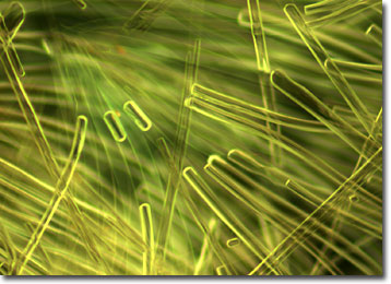
The specimen presented here was imaged with a Nikon Eclipse E600 microscope operating with fluorite and/or apochromatic objectives and vertical illuminator equipped with a mercury arc lamp. Specimens were illuminated through Nikon dichromatic filter blocks containing interference filters and a dichroic mirror and imaged with standard epi-fluorescence techniques. The specific filter utilized for the milkweed fibers was a B-2E/C. Photomicrographs were captured with a Nikon DXM 1200 digital camera system coupled to the microscope with a lens-free C-mount adapter.
BACK TO THE FLUORESCENCE DIGITAL IMAGE GALLERY