
Fluorescence Digital Image Gallery
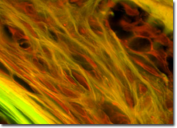
Mammalian Spongy Bone
There are two basic structural types of bone in mammals, compact and spongy. Spongy bone, also known as cancellous bone, is less dense than compact bone and is composed of a honeycomb-like network of bones called trabeculae. Spongy bone actually looks like a sponge. It is found at the expanded heads of long bones, such as bones of the arms, legs, fingers and toes.
Spongy bone also fills most irregular bones, such as bones of the skull, vertebrae and hips. Blood vessels run throughout the substance and marrow fills the spaces between the tissues. Spongy bones are designed to bear stress caused by bending or stretching.
Bones are major components of the skeletal systems of nearly all vertebrate animals. There are 206 bones in the adult human, which make up 14 percent of total body weight. Bone is composed of both living and nonliving materials and provides a framework for the body and a surface for the attachment of muscles. The skull and rib bones protect the brain and heart. Bones are also a vital part of the body's physiology. They store calcium, which is essential for nerve and muscle cells. Also, the soft core of bone, the bone marrow, is where red blood cells, certain white blood cells, and blood platelets form.
The specimen presented here was imaged with a Nikon Eclipse E600 microscope operating with fluorite and/or apochromatic objectives and vertical illuminator equipped with a mercury arc lamp. Specimens were illuminated through Nikon dichromatic filter blocks containing interference filters and a dichroic mirror and imaged with standard epi-fluorescence techniques. Specific filters for the mammalian spongy bone specimen were a UV-2E/C, B-2E/C, and a Y-2E/C. Photomicrographs were captured with an Optronics MagnaFire digital camera system coupled to the microscope with a lens-free C-mount adapter.
BACK TO THE FLUORESCENCE DIGITAL IMAGE GALLERY