
Fluorescence Digital Image Gallery
Mammalian Compact Bone
There are two basic structural types of bone in mammals, compact and spongy. Compact bone is very dense and hard on the outside, and makes up most of the bones in the arms and legs. The structural units are osteons, which are elongated cylinders acting as weight-bearing pillars that can withstand high levels of mechanical stress.
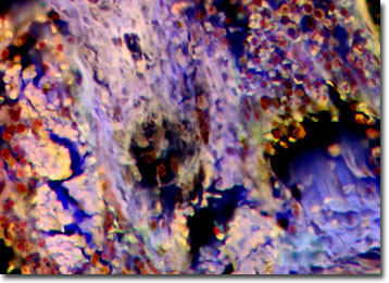
The osteons' center is a hollow canal that acts as a central passageway for blood vessels and nerves. A thin membrane, termed the periosteum, surrounds compact bone. The outer layer of this membrane contains blood vessels and nerves that travel into the bone. The inner layer consists mainly of osteoblasts, cells that are constantly renewed in the bone. Osteoblasts make and secrete collagen, giving bone a measure of elasticity. Also, osteoblasts secrete mineral salts formed from calcium and phosphorus, which prevent the bones from breaking easily, as well as rebuilding bone if necessary.
Bones are major components of skeletal systems in nearly all vertebrate animals. There are 206 bones in the adult human, which make up 14 percent of total body weight. Bone is composed of both living and nonliving materials and provides a framework for the body and a surface for the attachment of muscles. The skull and rib bones protect the brain and heart. Bones are also a vital part of the body's physiology. They store calcium, which is essential for nerve and muscle cells. Also, the soft core of bone, the bone marrow, is where red blood cells, certain white cells and blood platelets form.
The specimen presented here was imaged with a Nikon Eclipse E600 microscope operating with fluorite and/or apochromatic objectives and vertical illuminator equipped with a mercury arc lamp. Specimens were illuminated through Nikon dichromatic filter blocks containing interference filters and a dichroic mirror and imaged with standard epi-fluorescence techniques. Specific filters for the mammalian compact bone specimen were a UV-2E/C, B-2E/C, and a Y-2E/C. Photomicrographs were captured with an Optronics MagnaFire digital camera system coupled to the microscope with a lens-free C-mount adapter.
BACK TO THE FLUORESCENCE DIGITAL IMAGE GALLERY