
Fluorescence Digital Image Gallery
Lone Star Tick
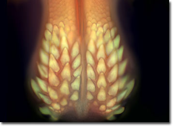
Amblyomma americanum is commonly known as the lone star tick. It is named for the dramatic iridescent spot that can be found on the female. Lone star ticks are found primarily in northern and central Florida, but can also be found in the mid-Atlantic and south-central parts of the United States, as well as Mexico.
Humans, pets, livestock and wildlife are all potential hosts for the lone star tick. This species is known to transmit canine ehrlichiosis, human monocytic ehrlichiosis, and granulocytic ehrlichiosis; diseases that attack the white blood cells. In most cases, infections are mild or without symptoms, but without proper diagnosis and treatment the resulting disease can be severe and even life threatening. Ehrlichiosis disease resembles the Rocky Mountain spotted fever. Common symptoms include sudden high fever, fatigue, muscle aches, severe headache, and sometimes rashes. Lone star ticks are also reported to transmit Rocky Mountain spotted fever, Q fever, tularemia, and Lyme disease.
The specimen presented here was imaged with a Nikon Eclipse E600 microscope operating with fluorite and/or apochromatic objectives and vertical illuminator equipped with a mercury arc lamp. Specimens were illuminated through Nikon dichromatic filter blocks containing interference filters and a dichroic mirror and imaged with standard epi-fluorescence techniques. The filter combination utilized for the lone star tick was a triple cube specific for DAPI, FITC, and Texas Red. Photomicrographs were captured with an Optronics MagnaFire digital camera system coupled to the microscope with a lens-free C-mount adapter.
BACK TO THE FLUORESCENCE DIGITAL IMAGE GALLERY