
Fluorescence Digital Image Gallery
Human Hyaline Cartilage
Cartilage is a dense network of collagen fibers, embedded in a firm but plastic-like gelatinous substance covered by a membrane called the perichondrium. Mammals have three types of cartilage: hyaline, elastic, and fibrocartilage.
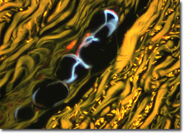
The type of protein fiber in the cartilage matrix determines its type. In hyaline, the protein fibers are mostly collagen, which provides flexibility and support at the joints. Hyaline contains very little elastin compared to elastic cartilage. Hyaline cartilage is the most widely distributed form and is also the type that makes up the embryonic skeleton. In adults, it's found at the ends of bones in free-moving joints and at the ends of the ribs, and also in the nose, larynx, trachea and bronchi. It is pearly, bluish-gray and semitranslucent in appearance and very resilient. Articular cartilage is a type of hyaline that is made up of thin, transparent sections easily cut by a knife. Cartilage is found covering the articular surfaces of bones and synovial joints. Articular cartilage is deformable but quickly recovers its shape, which is an important characteristic for its functions of reducing friction and absorbing shock.
The primary function of cartilage in mammals is to form a model for later growth of the bony skeleton, but some parts of the skeleton will retain the cartilage form into adulthood. Unlike bone, cartilage is avascular, i.e. it contains no blood vessels. Specialized cells, called chondrocytes, are distributed throughout the collagen gel matrix and obtain nutrition by diffusion through the gel.
The specimen presented here was imaged with a Nikon Eclipse E600 microscope operating with fluorite and/or apochromatic objectives and vertical illuminator equipped with a mercury arc lamp. Specimens were illuminated through Nikon dichromatic filter blocks containing interference filters and a dichroic mirror and imaged with standard epi-fluorescence techniques. The specific filter for the human hyaline cartilage stained thin section was a DAPI, FITC, Texas Red combination. Photomicrographs were captured with an Optronics MagnaFire digital camera system coupled to the microscope with a lens-free C-mount adapter.
BACK TO THE FLUORESCENCE DIGITAL IMAGE GALLERY