
Fluorescence Digital Image Gallery
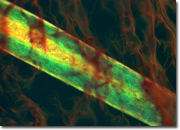
Human Scalp
In humans, the scalp is a specialized area of skin on top of the head, usually covered with hair in both sexes. It consists of three layers: a layer of skin, an underlying layer of tissue and blood vessels, and the occipitofrontalis muscle, which raises the eyebrows, stretching from the top of the eyebrows to the back of the head. The scalp covers most of the head, starting at the top of the forehead, and contains as many as 150,000 hair follicles.
Scalp itchiness is a problem afflicting 43 percent of the general population at least once in a 12-month period. Many factors contribute to this including blow dryers, styling products, wool hats, allergies, dandruff, mites, and even stress. The scalp can reflect the overall condition of the body and is affected by stress and hormonal changes. A overstressed immune system lowers resistance, which can result in over-colonization of a yeast organism called Pityrosporum ovale. Although it's a normal part of the human skin flora, an overpopulation of this fungus can cause dermatitis and dandruff.
Aging causes changes in the human scalp, such as the gradual thinning and graying of hair. Receding hairlines and baldness are common in men. These changes affect the hair follicle and are a result of the individual's genetics and male sex hormones.
The specimen presented here was imaged with a Nikon Eclipse E600 microscope operating with fluorite and/or apochromatic objectives and vertical illuminator equipped with a mercury arc lamp. Specimens were illuminated through Nikon dichromatic filter blocks containing interference filters and a dichroic mirror and imaged with standard epi-fluorescence techniques. The specific filter utilized for the human scalp stained thin section was a B-2E/C. Photomicrographs were captured with a Nikon DXM 1200 digital camera system coupled to the microscope with a lens-free C-mount adapter.
BACK TO THE FLUORESCENCE DIGITAL IMAGE GALLERY