
Fluorescence Digital Image Gallery
Human Roundworm
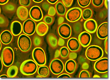
Ascaris lumbricoides, the large human roundworm, is the most massive nematode to parasitize humans, growing up to 16 inches long and often as thick as a pencil. Infections of this intestinal roundworm, called ascariasis, are extremely common in rural communities around the world and affect as many as 1.5 billion people, almost one-quarter of the world's population.
Infections occur after people ingest food or soil contaminated with Ascaris eggs. Unlike many other parasites, infections are direct and do not require a secondary host. An almost identical worm, Ascaris suum, occurs in pigs, but is physically indistinguishable from Ascaris lumbricoides.
After the eggs of the human roundworm hatch in the small intestine, larvae are liberated and migrate through the intestinal wall. From there, they enter the blood stream and travel to the lungs, where they may produce lung inflammation and fluid retention, or even severe hemorrhagic pneumonia. About 10 days later, the larvae migrate up through the air passages of the lungs, to the trachea. They enter the throat and are swallowed, eventually ending up in the small intestine where they mature and mate, completing their lifecycle. Serious, potentially fatal, complications of ascariasis can result when larvae lodge in sensitive tissues, such as the brain, and when adult worms migrate into various body structures where they produce abscesses and toxic manifestations. Intestinal blockages from adult worms sometimes require surgical removal.
The specimen presented here was imaged with a Nikon Eclipse E600 microscope operating with fluorite and/or apochromatic objectives and vertical illuminator equipped with a mercury arc lamp. Specimens were illuminated through Nikon dichromatic filter blocks containing interference filters and a dichroic mirror and imaged with standard epi-fluorescence techniques. Specific filters for the roundworm stained thin section were a B-2E/C and a Y-2E/C. Photomicrographs were captured with an Optronics MagnaFire digital camera system coupled to the microscope with a lens-free C-mount adapter.
BACK TO THE FLUORESCENCE DIGITAL IMAGE GALLERY