Fluorescence Digital Image Gallery
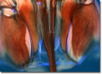
Honeybee Stinger
The honeybee's stinger is smaller than the head of a pin, but its venom can produce pain worse than a hypodermic needle. It's even worse for the two of every one hundred people who are allergic to bee venom, resulting in swelling, rash, dizziness, and even anaphylactic shock.

The stinger is located on female bees at the end of the abdomen and is part of the ovipositor, which is an egg-laying device. Even though most bees can sting repeatedly, a honeybee only has one chance. The honeybee stinger has a hook-shaped barb and when it catches in a victim the bee can't fly away without inflicting the fatal wound of tearing out its ovipositor along with some internal organs. Even after the bee detaches itself, the venom sac and its attached muscles continue to pump venom into the victim.
Concerns about bee stings recently entered the public health arena with the spread of killer bees throughout South and North America. In 1957, African bees were imported into Brazil for breeding experiments, but escaped and mated with previously imported European honeybees. The Africanized honeybees established themselves as a particularly aggressive breed and soon earned themselves the name "killer bees." Swarms of Africanized honeybees have been known to kill small and large farm animals as well as dozens of humans. While their venom is no more potent than that of the European honeybee, the Africanized honeybees react more quickly to a threat, attack in greater numbers, pursue for a longer time, and take longer--as long as 24 hours--to calm down. Africanized honeybees have been spreading north over the decades, through Argentina and northward throughout South and Central America, and Mexico. They first entered the United States in Southern Texas in 1990. Since then, colonies have been found in California, New Mexico, Nevada, and Arizona.
The specimen presented here was imaged with a Nikon Eclipse E600 microscope operating with fluorite and/or apochromatic objectives and vertical illuminator equipped with a mercury arc lamp. Specimens were illuminated through Nikon dichromatic filter blocks containing interference filters and a dichroic mirror and imaged with standard epi-fluorescence techniques. Specific filters for the bee stinger were a UV-2E/C and a Y-2E/C. Photomicrographs were captured with a Nikon DXM 1200 digital camera system coupled to the microscope with a lens-free C-mount adapter.
BACK TO THE FLUORESCENCE DIGITAL IMAGE GALLERY
