
Fluorescence Digital Image Gallery
Honeybee Leg
Honeybees are classified in any of the four species that belong to the genus Apis. They are classified, along with 20,000 other insect species, as members of the Apoidea superfamily (order Hymenoptera), which includes ants, wasps, hornets, and many other species of bees.
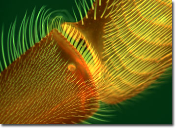
A honeybee's body is divided into three segments, each one equipped with a pair of legs. Each pair of legs is highly specialized and, in combination, they make the perfect tools for collecting pollen, which provides essential proteins for honeybee larvae. The front legs and feelers are used for tasting and removing pollen that has been collected on the front of the body and head. The middle legs remove the pollen from the first pair of legs and the thorax with the flat, wide, hairy tarsal joint, which acts like a brush. On the lower end of the middle legs is a spine, called the spur, which removes plates of wax from the abdomen. The hind legs carry the pollen load.
Flowers tend to lend a helping hand to honeybees. The pollen grains of flowers most attractive to the bees are very sticky so the pollen will secure easily to their bodies. Also, many flowers provide a landing lip for the bees to sit on before diving into the flower's interior. Larkspur, monkshood, bleeding heart, and Scotch broom are some of the flowers that depend on bees for pollination. A large honeybee colony may collect over 1,000 pounds of nectar and pollen in one year, hence the idiom, "As busy as a bee."
The specimen presented here was imaged with a Nikon Eclipse E600 microscope operating with fluorite and/or apochromatic objectives and vertical illuminator equipped with a mercury arc lamp. Specimens were illuminated through Nikon dichromatic filter blocks containing interference filters and a dichroic mirror and imaged with standard epi-fluorescence techniques. The filter combination utilized for the honeybee leg was a triple cube specific for DAPI, FITC, and Texas Red. Photomicrographs were captured with an Optronics MagnaFire digital camera system coupled to the microscope with a lens-free C-mount adapter.
BACK TO THE FLUORESCENCE DIGITAL IMAGE GALLERY