
Fluorescence Microscopy
Combination Methods with Phase Contrast
To minimize the effects of photobleaching, fluorescence microscopy can be combined with other techniques that are non-destructive to the fluorochrome, such as differential interference contrast (DIC), Hoffman modulation contrast (HMC), transmitted darkfield illumination, and phase contrast.
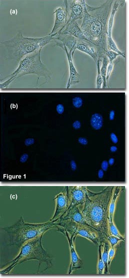
The idea is to locate a specific area of interest in a specimen using the non-destructive contrast enhancing technique then, without relocating the specimen, switch the microscope to fluorescence mode. The results of a typical experiment of this type are illustrated in Figure 1. Figure 1(a) illustrates 3T3 fibroblasts in monolayer tissue culture imaged using phase contrast optics. The cell line was established from a National Institutes of Health line of Swiss mouse embryo cells, which are highly contact inhibited and useful for studies involving sarcoma virus formation and leukemia virus propagation. The photomicrograph in Figure 1(b) shows the same viewfield, but this time imaged using fluorescence illumination (a mercury vapor lamp and an Olympus WU filter cube) with cells stained by the fluorochrome 4',6-diamidino-2-phenylindole (DAPI), a nucleic acid specific dye with an emission maximum at 461 nanometers, which is used to selectively stain nuclei and chromatin. Figure 1(c) illustrates the two techniques used in combination to produce a beautiful photomicrograph of fluorescent-stained 3T3 cellular nuclei superimposed on a phase contrast image of the fibroblast cell membranes and internal organelles. This image was recorded with a specialized fluorite objective designed with phase rings to permit simultaneous observation of both fluorescence and phase contrast with the same objective.
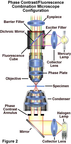
The microscope configuration typically utilized to simultaneously image specimens using both phase contrast and fluorescence illumination is illustrated in Figure 2. Phase contrast is conducted using transmitted light provided by a tungsten-halogen lamp positioned in a lamphouse attached to the microscope base. Light passing through the field diaphragm is reflected by a mirror into the substage condenser and through a phase annulus of the proper dimensions.
| Interactive Tutorial | |||||||||||
|
|||||||||||
Light diffracted by the specimen is first passed through a phase plate positioned in the objective rear focal plane before interfering at the intermediate image plane with light passing through the specimen undiffracted. Simultaneously, ultraviolet light emitted by a mercury burner is passed through an exciter filter, then reflected by a dichroic mirror onto the specimen from above. Secondary fluorescence emitted by the chromophore attached to the stained specimen is captured by the objective and passed through the barrier filter and into the eyepieces and/or phototube. This configuration can be used to image specimens using the techniques (fluorescence and phase contrast) individually or in combination.
Contributing Authors
Mortimer Abramowitz - Olympus America, Inc., Two Corporate Center Drive., Melville, New York, 11747.
Michael W. Davidson - National High Magnetic Field Laboratory, 1800 East Paul Dirac Dr., The Florida State University, Tallahassee, Florida, 32310.
BACK TO FLUORESCENCE MICROSCOPY
BACK TO PHASE CONTRAST MICROSCOPY