Differential Interference Contrast Image Gallery
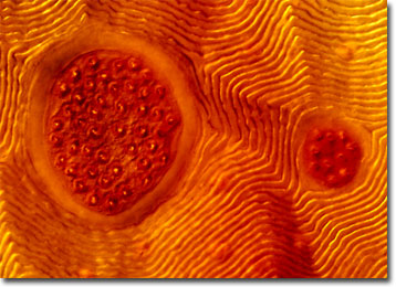
Rocky Mountain Wood Tick
Scientifically described as Dermacentor andersoni, the Rocky Mountain wood tick gains its common name from its primary habitat in the United States, although the species can also be found in southwestern Canada. The bloodsucking parasite is one of the two major vectors of the typhus-like disease known as Rocky Mountain spotted fever.

The lifecycle of the Rocky Mountain spotted wood tick generally takes two to three years to be completed and requires the parasitism of three different hosts. After each blood meal, the tick drops to the ground and molts into its subsequent form, or dies if it has already successfully completed the mating process. To find an appropriate host, the Rocky Mountain spotted wood tick climbs grass, bushes, and other foliage, where it waits for chemical cues that indicate the presence of a mammal. The parasite then drops itself onto the mammal and briefly moves about before attaching its mouthparts to the hostís skin.
In its larval and nymph forms, the Rocky Mountain spotted wood tick feeds on small rodents from which it may become infected with Rickettsia rickettsii. The bacteria can then be passed on to larger creatures, such as humans, that the tick feeds upon in its adult stage, resulting in the spread of Rocky Mountain spotted fever. Symptoms of the disease typically include a red rash and a fever, but may lead to paralysis, deafness, gangrene, or even death in less than a week. Early treatment with antibiotics greatly decreases the possibility of the diseaseís most serious consequences.
