
Differential Interference Contrast Image Gallery
Planaria Cross Section
Planaria are primarily free-living flatworms that are members of the class Turbellaria. Observers can go “fishing” for the invertebrates, which frequent the sediments of ponds and lakes, by simply tossing a piece of liver attached to a string into the water.
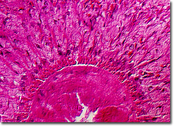
Arrow-shaped heads, paired ocelli, or eyespots, and long, flat bodies, make planaria fairly easy to recognize. In tropical regions the flatworms may be brightly colored, but North American species are usually brown, gray, or black. Planaria normally grow to be only a fraction of an inch long, but some may achieve lengths of more than a foot. Their tapered forms are edged in hair-like cilia, which help propel them forward.
Hermaphroditic, planaria may reproduce asexually by regeneration or by exchanging sexual gametes with another worm. Scientific evidence suggests if reproduction is asexual, then certain conditioned responses can be passed on to offspring. For instance, although they possess primitive central nervous systems, planaria are able to learn to travel through a maze to eat or react to other simple stimuli. After regeneration, these learned actions are remembered by the parent planaria, but are also adopted by the newly formed turbellarian.
Planaria are generally carnivorous night feeders that consume aquatic insects, snails, microcrustaceans, and proteinaceous detritus, though a few species are parasitic. The mouths of the flatworms are located on their ventral side more than halfway toward the tail, an opening through which they may extend the pharynx. Typically, enzymes secreted by the mouth partially digest prey while it is pinned outside the body by the pharynx. Once softened, the mouth sucks in the food and digestion continues within a three-branched gastrovascular cavity.