Differential Interference Contrast Image Gallery
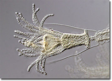
Obelia Hydroid
Obelia is a genus of invertebrate marine organisms whose members exist in alternate generations as polyps and medusae. Part of the phylum Cnidaria, Obelia species are related to sea anemones, corals, freshwater hydra, and jellyfish.

The polyp members of Obelia are asexual, stalk-like, and usually attached to the ocean floor, rocks, shells, or other surfaces. The polyps generate additional polyps by budding, creating a branching colony of the organisms that has a structure similar to that of a tree. A transparent sheath known as the perisarc encases the colony. Dimorphic, some of the polyps are responsible for feeding, while others concentrate their energy on reproduction. The feeding polyps feature tentacles and share the nutrients they imbibe with the rest of the colony after digestion in the gastrovascular cavity. Reproductive polyps, however, lack tentacles and are club-shaped.
The following generation of Obelia organisms are produced by the reproductive polyps via budding and are free-swimming. Similar in appearance to umbrellas, the medusae exhibit stinging tentacles that hang downward in the water. Inside the organisms are interconnected passageways that enable the distribution of water, nutrients, and oxygen, as well as waste removal. The medusae reproduce sexually, eggs and sperm combining in the surrounding water to develop a zygote. After growing into a planula larva, the organism finds a place to settle and transforms into a polyp that soon creates its own colony.
