
Differential Interference Contrast Image Gallery
Diatom Frustule
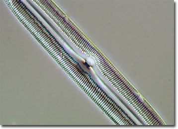
A frustule is the pillbox-like silica shell that encases a diatom. The various delicate and intricate designs that are characteristic of frustules make them particularly useful in microscopy, where they can be used to test the resolving power of an instrument.
Diatoms are unicellular phytoplankton that may be found in all of the Earth's aquatic environments. However, most species occur solely in areas that meet specific physical, chemical, and biological requirements. This habitat specificity is frequently utilized by ecologists to ascertain the state and quality of a body of water. Furthermore, the extensive fossil record of diatoms provides scientists with a unique historic record of changes in marine ecosystems.
Since frustules are unable to grow, the reproduction of diatoms is a curious and unusual process. The tiny organisms primarily proliferate asexually by cell division. In such instances, the overlapping halves of the frustule separate and each secretes a smaller lower half. Thus, one of the pair of diatoms remains approximately the size of the mother cell, while the other, which developed from the lower half of the original diatom, is smaller. Asexual reproduction dominates until a point is reached where the second cell cannot get any smaller, at which time sexual reproduction occurs in order to restore the cell line to its original dimensions.