Differential Interference Contrast Image Gallery
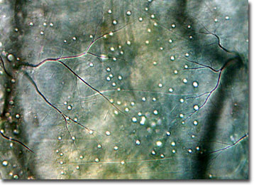
Chironomid Fly Larva
The general public often confuses chironomids, more commonly known as nonbiting midges, with a variety of biting flies and mosquitoes. These members of the order Diptera are, however, harmless and are important elements in the food chain, providing nutrients to many freshwater fish and migratory shorebird species.

The metamorphic lifecycle of a chironomid consists of several phases, including egg, larval, pupal, imago, and flying adult stages. It is only in their larval form that the flies can feed, so during that time they must consume enough nutrients to last them the rest of their lives. Worm-like in appearance, chironomid larvae typically inhabit the sediments of freshwater bodies where they feed upon algae and other small organisms. They may also exist, however, in marine or terrestrial environments. One unusual species, aptly nicknamed the ice worm, actually proliferates in the icy cold waters of New Zealandís west coast glaciers.
In certain ecosystems, such as the 41,000-acre Cheyenne Bottoms wetlands in Kansas, chironomid larvae are particularly abundant. As many as 50 of the creatures may inhabit a single square inch of mud, providing ample fuel for the large numbers of birds that visit the area during spring migration. Yet, some locales are not so fortunate. Aerially sprayed insecticides eliminate large numbers of chironomids, as well as mosquitoes and other disease-vector insects at which they are targeted.
